Nikon Ti2-E Confocal Inverted Microscope
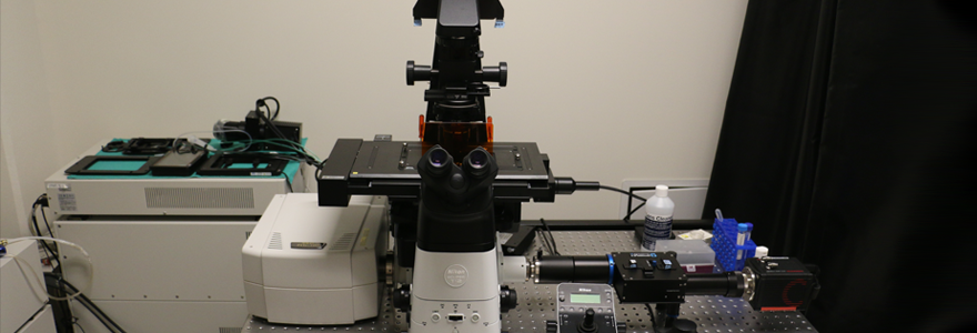
The Nikon Ti2-E Inverted Microscope paired with the A1R HD laser-scanning confocal offers an 18 mm field of view; which means more data per image, less stitched images and fewer artifacts. With its real-time hardware focus correction and intuitive/user-friendly operation, the A1R HD is perfect for imaging both fixed tissue, live cell, and intravital preparations.
Apart from live tissue imaging, the Nikon laser scanning confocal microscope can be used for imaging of slides and cell culture assays with improved results. Live cell imaging is possible using an incubator with temperature, CO2 and humidity control. Images of fixed tissues can be obtained in high resolution.
There are six lasers available for use with this microscope covering DAPI, CFP, FITC, YFP, TRITC, and CY5 labels. Objectives that are available include 10x air, 20x air, 20x water, 40x oil and 60x oil. The Nikon A1R HD point scanner is also capable of imaging in transmittance mode (DIC) without the use of labels.
Furthermore, interference filters and a 80-20 split mirror enable measurement of RBC oxygen saturation and flow parameters in microvasculature using absorption spectroscopy. Alpha Mass Flow Controllers allow changes in oxygen levels in a custom-chamber under the intravital preparation to challenge microvasculature. Responses to such changes are measured via changes in flow or with perictyes in the tissue.
Nikon Elements denoise.ai Software
Key Features
- A high performance laser unit that offers six wavelengths: 405, 445, 488, 514, 561, and 640 nm.
- Six objectives: 4X, 10X, 20X, 20X WI, 40X oil, and 60X oil.
- A high-speed resonant scanner that allows imaging of intracellular dynamics at 30 fps (512 x 512 pixels) and 420 fps (512 x 32 pixels).
- A Galvano scanner with high-speed acquisition capability of 10 fps (512x512) and 130 fps (512x32) and high-resolution of imaging up to 4096x4096 pixels.
- Perfect Focus System (PFS) which continuously corrects for focus drift during time-lapse observations.
- A Detector unit—comprised of two PMTs and two GaAsP (gallium arsenide phosphide) detectors—which is significantly more sensitive than conventional PMTs.
- Optosplit II Bypass with a Hamamatsu Orca Flash 4 camera for brightfield or absorption spectroscopy.
-
Live cell imaging chamber
- High speed piezo drive for Z-stacking and large tiled images
- Nikon Elements software
Gallery
In vivo 3D scan of a mouse skeletal muscle venule after intravenous injection of a plasma tracer (green; FITC-dextran) and leukocyte stain (red; rhodamine 6G). Captured on Nikon Ti2-E Confocal Inverted Microscope by Stephanie Milkovich and Paulina Kowalewska.
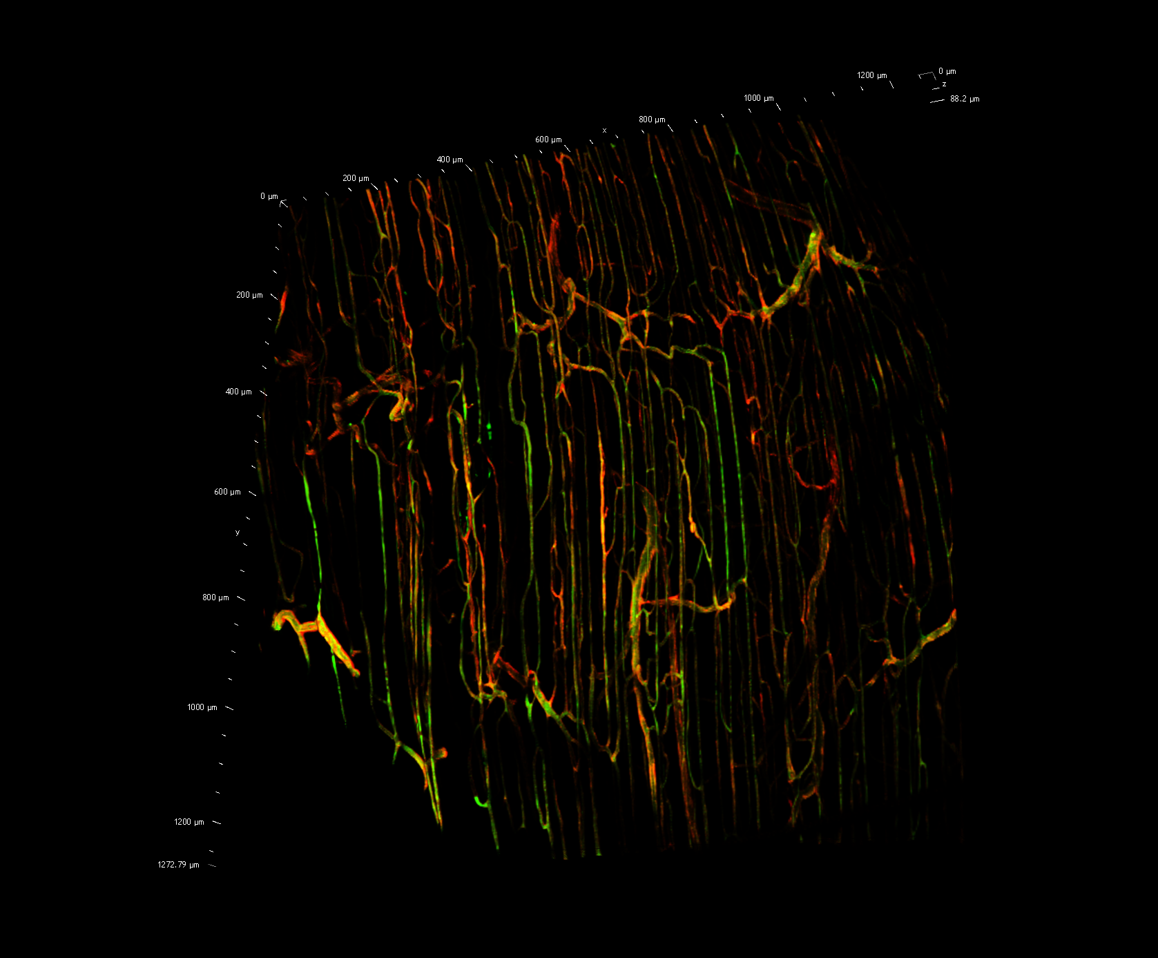
Stitched in vivo image of the mouse skeletal muscle microvasculature with FITC-dextran plasma tracer (green) and CD31-labeled endothelium (red). Captured on Nikon Ti2-E Confocal Inverted Microscope by Stephanie Milkovich and Paulina Kowalewska.


